Arteriovenous Malformation
An arteriovenous malformation (AVM) is a congenital tangle of dilated blood vessels in which arterial blood flows abnormally and directly from arteries into draining veins. These so-called “hot” shunts within the vascular tangle lead to the formation of bulges in the vessel walls and, over time, to sudden bleeding of varying extent. Bleeding may recur, and depending on its location, the hemorrhage may cause impairment of brain function, whether focal or global, especially if blood enters the cerebrospinal fluid in large amounts. This can also disrupt cerebrospinal fluid circulation, which requires early neurosurgical intervention.
In addition to these relatively dangerous vascular malformations, CT and MRI may incidentally reveal much more common, less dangerous vascular malformations. These include developmental venous anomalies and cavernomas (and, very rarely, telangiectasias). Developmental venous anomalies are abnormal venous drainage patterns and are usually followed conservatively, while cavernomas may cause epilepsy and may also be multiple.
What Causes Cerebral AVMs?
AVMs are a manifestation of a group of disorders involving an abnormality of the vascular endothelium, the inner lining of blood vessels. In most patients, there is only an inherited predisposition to their development. A genetic basis has been identified in only a fraction of patients, typically those with a family history, such as hereditary hemorrhagic telangiectasia, Wyburn-Mason syndrome, or RASA1 mutations. AVMs cannot be prevented, but once diagnosed, they require appropriate treatment and follow-up.
What Symptoms Can Occur With AVM?
Chronic headaches are highly nonspecific and common in the general population.
Epileptic seizures may lead to the diagnosis of AVM through MRI, which is part of the standard workup for new-onset epilepsy.
Brain or cerebrospinal fluid hemorrhage of varying severity, sudden headache, paralysis, and impaired consciousness can all lead to a diagnosis.
How Is Cerebral AVM Treated?
Approach depends on the symptoms. AVMs that have bled are treated to prevent further hemorrhage, remove the hematoma, and resect the AVM itself.
Treatment is timed according to life-threatening symptoms. If the hematoma is small and neurological deficits are mild, neurosurgery may be performed after appropriate preparation within days.
For unruptured brain AVMs, radiosurgery with a Leksell Gamma Knife is most often considered.
Transcatheter endovascular occlusion is a procedure that follows diagnostic angiography. It may be combined, most often with microsurgical resection or with irradiation, in either order.
Treatment and follow-up of patients with cerebral AVMs are provided in comprehensive cerebrovascular centers, and follow-up may also be coordinated with an outpatient neurologist.
What Can I Do Myself?
After diagnosis, it is important to follow the recommended check-ups.
Epileptic seizures require regular medication to prevent recurrence.
Brain AVMs can be small (roughly up to 3 cm) and relatively easy to treat surgically if, for example, they are located on the brain surface. Deeply located AVMs are often better treated with stereotactic radiosurgery. There are also extensive AVMs in which invasive treatment does not change the unfavorable course, and in such cases we proceed individually, now with the possibility of influencing the disease using so-called biological therapy.
AVMs in children are divided into those that are truly congenital (present at birth) and AVMs occurring in juvenile age. Pediatric AVMs require specialized care. They are generally rare, and if they present with bleeding, they tend to be aggressive, with a need to exclude recurrence after treatment.
Spinal AVMs require specialized, comprehensive care, including rehabilitation, and patients are referred to specialized centers, including centers within the EU.
Findings on MRI and CT reported simply as “vascular malformation” should be clarified as to the exact type, such as developmental venous anomaly or cavernoma, so that the patient is not unnecessarily stressed. Their presence may not require follow-up at all, or a control scan may be recommended after one year, for example.
Who Should I Contact?
Consultation for a diagnosed cerebral AVM belongs in a neurology and neurosurgery clinic, after which the patient is referred directly to a specialized center. It is usually necessary to discuss the treatment options in detail, including the risks of each treatment method, ideally in the presence of family members.
AVM Classification
Based on morphological structure, AVMs are divided into:
- AVMs with a compact (glomerular) nidus – typically abnormal vessels without brain tissue; this type is more common than the diffuse type.
- AVMs with a diffuse (proliferative) nidus – there is no well-formed nidus with functional neuronal tissue dispersed among the abnormal vessels.
S-M grading
In clinical practice, the classification known as the Spetzler-Martin grading system is used. It assigns points based on the size of the malformation, the type of venous drainage, and whether the AVM is located in an eloquent area of the brain. According to this classification, AVMs are divided into five grades:
- Low-grade – grade I, II and III (1–3 points),
- High-grade – grade IV and V (4–5 points).
Scoring table according to the S-M grading system:
The Spetzler-Martin grading system is used primarily for surgically resectable AVMs. It lacks many parameters that are fundamental for deciding on radiosurgical or endovascular treatment. Therefore, additional classifications have been developed for these modalities; for radiosurgery these include, for example, the Virginia grading system and the Pollock-Flickinger grading system. For endovascular treatment, the best known are the Feliciano grading system and the Buffalo score.
Angioarchitecture
An AVM usually has a well-defined center, or nidus, and dilated feeding arteries together with enlarged draining veins filled with arterialized blood. In this type of vascular malformation, the resistance of the capillary bed is absent, so flow is markedly increased.
Epidemiology
The incidence in the population is relatively low, with newly diagnosed cases reaching 1–1.5 per 100,000 inhabitants. AVMs most often present at a younger age, typically between 15 and 20 years of age.
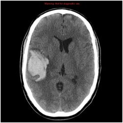
CT without contrast shows a large hematoma in the right temporoparietal region, caused by rupture of an AVM.
Location
CNS
In the brain, AVMs are located supratentorially in 86% of cases, with those in the neocortex being the most common, followed by deeply seated AVMs in the diencephalon, basal ganglia, internal capsule, and related areas, as well as combined AVMs in the archicortical and paleocortical regions. Infratentorial AVMs are uncommon and mainly affect the neocerebellum and brainstem,intraventricular AVMs have also been reported. Eighty percent of spinal AVMs are intramedullary or extramedullary.
Hemorrhage from AVMs is approximately 30% subarachnoid, 23% intraparenchymal, 16% intraventricular, and 31% a combination of the above.
Other Location
Other sites where AVMs may occur include:
- lungs,
- liver,
- kidneys,
- uterus and others
Diagnosis
CT
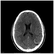
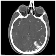
CT is often the first imaging method used in patients with acute neurological deficits, but without contrast, an accurate diagnosis of AVM is relatively difficult. The nidus of an AVM without prior hemorrhage is hyperdense relative to the surrounding brain tissue. After contrast administration, especially on CTA, the diagnosis becomes clearer, but angiography is recommended for detailed visualization of the feeding arteries. In the case of AVM rupture, CT is sufficiently sensitive to detect an acute hematoma.
CT shows the difference in AVM diagnosis with and without contrast administration.
MRI
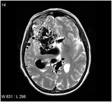
MRI – cerebral AVM
MRI is especially important for clearly showing adjacent structures and any associated damage. Neighboring edema is also well visualized. Compared with CT, MRI better defines the exact anatomical location of the nidus and any prior hemorrhage.
MRA
MRA sequences play an important role in producing three-dimensional angiographic images of AVMs. TOF MRA is typically one of the first examinations for such malformations and visualizes most feeding arteries and the nidus very well. However, it is often difficult to distinguish feeding arteries from venous drainage, especially in AVMs with more complex architecture. It is also not very sensitive for smaller AVMs and associated aneurysms.
Angiography – DSA
Digital subtraction angiography (DSA) is the gold standard for detailed AVM diagnosis. On angiography, the malformation appears as a tightly packed mass of enlarged surrounding arteries supplying the nidus. DSA provides a detailed description of the feeding arteries and associated angiopathic changes, precise evaluation of the nidus (its size, hemodynamic characteristics, and anatomical features), delineation of venous drainage (deep or superficial), and any signs of stenotic changes or ectasia. If hemorrhage has occurred, the hematoma typically compresses the nidus, thereby increasing intranidal vascular resistance. As a result, venous outflow may be delayed and the AVM may become angiographically occult: the malformation is anatomically present but not visible on angiography.
Associated Abnormalities
The most commonly observed abnormality associated with AVM is aneurysm, which typically develops as a result of increased blood flow through the malformation. Aneurysms may occur within the nidus (intranidal, present in up to 41–100% of AVM patients) or on feeding vessels (intrapedicular). In some cases, aneurysms unrelated to the nidus may also be seen on diagnostic imaging.
A flow-related angiopathy secondary to endothelial hyperplasia is also relatively common.
Clinical Presentation
The presentation of these vascular malformations is highly variable. Some AVMs may remain asymptomatic for a long time, while some patients may present with post-hemorrhagic neurological deficits, headaches, or epileptic seizures. Cardiac murmurs have also been reported in some patients.
A so-called steal syndrome is often seen: arteries supplying the AVM “steal” blood from surrounding vessels, which can result in ischemia of the areas whose blood supply has been diverted to the AVM.
Therapy
AVM treatment is a classic example of multimodal cooperation between several branches of medicine. AVMs may be surgically resected, treated with radiosurgery, or embolized endovascularly. In a single patient, several treatment options may be combined to achieve the best possible result with the lowest possible morbidity. Observation is, of course, a legitimate approach when the risk of rupture is not high and the malformation itself is not causing serious problems.
- Surgical resection is used especially for smaller, superficially located malformations. Its advantage is immediate elimination from the circulation and therefore definitive treatment. Complete resection is necessary, and resection may also eliminate an epileptogenic focus.
- Gamma Knife radiosurgery is useful for AVM nidi up to 2.5–3 cm in diameter and for deeply located lesions. Its advantage is that it is noninvasive, its disadvantage is that obliteration of the malformation occurs gradually over 1–3 years, during which the patient remains at risk of hemorrhage from the AVM.
- Endovascular embolization of the AVM nidus is rarely used as a standalone procedure. Its goal is to occlude deep feeding vessels before surgery or part of a large AVM nidus that was clearly the source of bleeding, or that could bleed, such as an intranidal aneurysm.
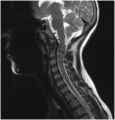
MRI − spinal AVM
Complications
In cases managed by observation, the greatest complication is hemorrhage. The risk of hemorrhage in patients without prior bleeding is approximately 2–3%. AVM hemorrhage, unlike cavernous malformations, can have fatal consequences because of its morphological structure; the literature reports post-hemorrhagic mortality of up to 30% and a 10–20% probability of persistent neurological deficits. In some cases, cerebral ischemia may occur, AVM rupture accounts for approximately 1–2% of all cerebral ischemic events. Aneurysms in the nidus region, or on feeding vessels, are also common and tend to rupture because of the high blood-flow velocity through the malformation.
Surgical resection is demanding and often takes several hours, but it can yield excellent long-term results when resection is radical. The aim is complete removal, which automatically eliminates the patient’s future risk of bleeding from that AVM. Some postoperative morbidity may occur, but in most patients it gradually improves over time; only a minority of patients, on average about 7%, have persistent morbidity.
Patients suitable for radiosurgical intervention should not have a high risk of hemorrhage, because the treatment effect does not appear immediately after irradiation. For a relatively long period, the patient remains at risk of bleeding from the AVM. One possible strategy is to irradiate the AVM first and operate afterward. Experience with this approach varies: some centers operate after radiosurgery only if irradiation was not sufficiently effective, while others use radiosurgery before surgical resection because the malformation usually bleeds less after irradiation.
The most common complication of endovascular embolization is again hemorrhage, for example due to perforation of an artery during microcatheter insertion or rupture of the AVM. There is also a risk of thrombus formation and migration to adjacent cerebral areas, which may result in ischemia.

